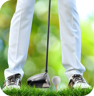
Golfer’s Elbow (Medial Epicondylitis) refers to the pain and inflammation that occurs at the inside point of the elbow (medial epicondylitis).
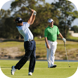
Golfer’s Elbow can be caused by any activity (not just golf) that requires forceful and repeated bending of the wrist and fingers. For example: when the golfer swings his club, the flexor muscles and tendons of the arm tighten just before the club makes contact with the ball. This repeated action stresses the muscles, causing micro- tearing of the flexor tendon, and inflammation of the soft-tissues. RSI problems occur when these muscles and tendons continue to be re-injured while the small tears are still in the process of healing. These new injuries cause the body to lay down additional adhesive scar tissue between the muscle layers in an attempt to stabilize the affected soft-tissues.
This adhesive scar tissue forms attachments to adjacent layers of tissue and structures, and inhibits the normal movement or translation of these soft-tissue structures. This lack of smooth movement causes friction and generates an ongoing cycle of inflammation and scar tissue formation.
Elbow injuries often require the practitioner to address a much bigger picture than just the elbow area. Symptomatically, what initially appears to be an elbow injury may actually be the result of poor core stability and lack of strength. This requires treatment of a much larger kinetic chain than just the elbow.
The Kinetic Chain of Golfer’s Elbow
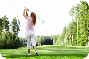
 The game of golf emphasizes one-sided activity of the body; you are either a right-handed golfer or a left-handed golfer. This unilateral focus is the cause of numerous injuries as golfers tend to develop muscle imbalances which cause a wide array of myofascial restrictions.
Golf, in its ideal form, is all about efficiently storing and releasing energy from your core out into your extremities. The classic golf swing engages your entire kinetic chain from your feet which form a solid stance, up through your hips and core, to finally release energy through your shoulders and arms right into the club head. This is much like a coiled spring, storing energy, then suddenly releasing it.
Unfortunately, for most golfers, this “coiled spring” is either broken or functions only minimally. Many golfers find that in the game of Golf, much of their energy and focus is spent on learning how to compensate for muscle imbalances, poor posture, and the multitude of myofascial restrictions that have developed over time.
Many patients who come to our clinic suffering from Golfer’s Elbow, also show these other common problems:
- Rounded shoulders (anterior posture).
- Restriction in the neck and low back (hypertonic or tight muscles with numerous trigger points).
- Tight restricted hips which are causing abnormalmotion patterns.
- Poor balance.
The Problem with Abnormal Motion Patterns
 A tight muscle on one side of your body almost always creates muscle imbalances, which in turn initiates a cascade of other events.
First, these muscle imbalances result in abnormal motion patterns, which then cause the body to compensate by using other structures to perform the required action.
These abnormal motion patterns result in increased friction between tissues layers, with the friction creating micro-tears in your muscles. The micro-tears induce increased inflammation and the eventual formation of scar-tissue.This scar tissue inhibits motion. Inhibitions in motion result in numerous additional compensations throughout the body, initiating yet another ongoing cycle of dysfunction.
In the case of a golfer, a restriction in the hips often causes the golfer to over-compensate with their shoulders during a golf swing. Over time, this over-compensation creates restrictions in that shoulder, along with a corresponding alteration in posture. This in turn leads to a cascading series of injuries and restrictions along the entire kinetic chain.
Tennis Elbow
 Tennis Elbow is a painful condition at the outside point of the elbow that typically involves inflammation and irritation of the extensor tendon where it attaches to the lateral epicondyle.
The injury process for Tennis Elbow (lateral epicondylitis) is identical to that of Golfer’s Elbow (medial epicondylitis). However, for Tennis Elbow, the pain manifests on the outside point of the elbow.
Tennis Elbow involves the extensors (the muscles that bend the wrist back). The extensors attach to the lateral epicondyle, on the outside of the elbow. The common extensor tendon also attaches to the lateral epicondyle. Both these structures are susceptible to micro-tears when they are exposed to repetitive actions.
As with Golfer’s Elbow, Tennis Elbow (Lateral Epicondylitis) can be caused by a variety of activities. Any activity that involves excessive supination (turning the hand, palm side up) and pronation, or lifting objects with your elbow in full extension (elbow straight) can cause this condition.
The repetitive motions of these activities result in the formation of micro-tears, inflammation, scar tissue, and physical restrictions which then manifest as Tennis Elbow. In addition to the layers of soft-tissue that surround the elbow, practitioners should also consider the numerous kinetic chain relationships when attempting to resolve this condition. Some of the affected structures are near the elbow while others are quite distant. Several layers of soft-tissues are typically involved in this injury, including:
- The deep annular ligament.
- The supinator and anconeus muscles.
- The superficial structures of the extensor muscles.
- The fascia that surrounds and penetrates through all these areas.
In most cases, both Golfer’s Elbow and Tennis Elbow can easily treated with Active Release Techniques. This is especially true if the practitioner applies a kinetic chain perspective to locate and treat ALL the affected structures.
The key point is to find all the affected structures and then treat each affected structure as needed. This type of treatment requires a high level of tactile sensitivity by the practitioner. Equally important for full recovery, the patient should carry out specially designed exercise routines that focus upon all the structures of the affected kinetic chain.
If you would like more information or to purchase our books please go to www.releaseyourbody.com.
If you would like information about our clinic in Calgary Alberta please go to www.kinetichealth.ca.
(COPYRIGHT KINETIC HEALTH 2012 – ALL RIGHTS RESERVED)
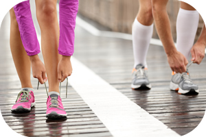
]]>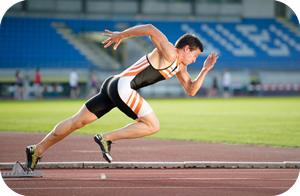 One of the most common running injuries we treat in our clinic is shin splints or Medial Tibial Stress Syndrome (MTSS). Shin splints cause one in five athletes to stop running. In addition to running, engaging in soccer, rugby, basketball, volleyball, or any sport that involves running or jumping can cause shin splints.
Symptoms
Most people would describe the initial pain of shin splints as a dull ache along the inside of the the lower leg (tibia). Shin splint pain is often felt at the beginning of a run and then diminishes as the run continues, only to return near the end of the run. In this initial stage the pain from shin splints will often dissipate completely with rest. If the shin splints progress, the pain will often be present with both activity and rest. Once shin splints reach the stage of constant pain, a medical professional should be seen to determine if additional injuries are present (stress fractures or compression syndrome).
The exact location of shin splints is often hard to find, because it is usually more of a diffused pain in the soft tissue (fascia, tendon, muscle) rather than on the bone (tibia) itself.
Causes of Shin Splints
 The most common cause of shin splints is repetitive motion. This is not surprising considering the force of impact of each runner’s stride. A runner’s shins are subject to two to three times the runner’s body weight on foot impact. This high level of force can easily overwhelm the shin muscles (dorsi flexors) if they are not strong.
Shin muscles are called dorsi flexors because of the motion they perform with contraction. On contraction the dorsi flexors pull the foot up towards your shins, this is what is referred to as dorsi flexion. During running, the dorsi flexor muscles also control plantar flexion, through the process of eccentric contraction. Eccentric contraction occurs when a muscle elongates while under tension. Controlling plantar flexion of the foot is not an exclusively linear motion. As the foot strikes the ground it is subjected to both rotational forces (pronation and supination) and side to side motions.
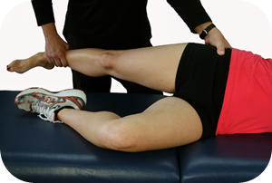
Any type of muscle imbalance, or abnormal motion pattern in the ankle, knee or hip could cause increased stress on the dorsi flexors as they try to control motion. In other words, the dorsi flexorsmay be the site of the shin splints, but the root cause could be far from the location of symptoms. For example, we know that excessive pronation (flat feet) will increase the load placed on thedorsi flexors as they try to control plantar flexion. We also know that weak or imbalanced external hip rotators (gluteus muscles) will increase pronation of the foot. Though it may not be a direct connection, weak external hip rotators could be part of the development and continuation of shin splints.
When it comes to shin splints, it is important to address the problems in all potential problem areas as well as the symptomatic structures in order to identify the root cause of the problem.
Differential Diagnosis
 It is very important in diagnosing shin splints to make sure that you are actually dealing with shin splints and not a stress fracture or a case of compartment syndrome.
Stress Fractures: See our page on Stress Fractures to review this condition.
Compartment Syndrome: Another condition that must be ruled out is compartment syndrome (CS). The muscles of our legs are divided into rigid compartments. These compartments are bound by strong fibrous tissue (deep fascia), and bones.
The anterior compartment contains some very important structures. It contains the dorsi flexors, the muscles directly linked to shin splints.
Anterior compartment Contains:
- Dorsiflexion muscles of the ankle and foot
- Tibialis anterior
- Extensor digitorum longus
- Extensor hallucis longus
- Peroneus tertius
- Anterior tibial artery
- Deep peroneal nerve
CS occurs when the pressure inside these compartments increases to the point where it interferes with the blood supply to your muscles and nerves. This can occur when the muscle inside the compartment becomes too large, increasing the pressure. CS can also occur from trauma, bleeding, swelling, overuse or even excessive medication.
In a case of anterior compartment syndrome, a runner may experience sharp pain and swelling over the shins. They may also notice weakness of the dorsi flexors, especially against resistance. In addition there is often a decrease in the extremities pulse and a decrease in sensation. There are two types of compartment syndromes: chronic and acute.
Chronic Compartment Syndrome is not a medical emergency and can often be treated with manual therapies (ART, Graston, Massage). Chronic CS is also referred to as exertional CS. The pain of exertional CS in runners usually comes on with the first 15 minutes of running, then subsides within an hour after the run. The pressure of these compartments can be measured by a medical practitioner. A resting pressure of greater than or equal to 15 mm Hg is an indication that compartment syndrome is present.
Acute Compartment Syndrome could be a serious limb-threatening condition. Any delay in treatment may lead to infection, complications and even limb amputation. In most cases an acute compartment syndrome occurs after a traumatic event, and is most commonly seen with traumatic fractures.
If you suspect a stress fracture or compartment syndrome you need the help of a medicial practitioner.
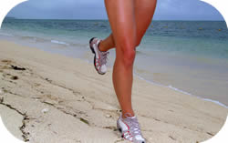
Running and Dorsi Flexor Strength
When a runner has weak dorsi flexors they will have a tendency to slap the ground with every foot strike. A runner slaps the ground because they are unable to control foot motion as they hit the ground (eccentric contraction). Next time you are out running, listen to the runners around you. You will be surprised to hear just how many of those runners are slapping the ground with their feet. These runners are susceptible to shin splints (MTSS).Another point to consider is that slow runners have a tendency to slap the ground more. In other words, they have weak dorsi flexors. This is a very interesting observation seeing that faster/elite runners are hitting the ground with more force, yet the fast runner is a quieter runner. This is because most elite runners have strong dorsi flexor muscles which are able to control the foot as it comes down. Essentially they have good shock absorbers that are able to dissipate the impact of each stride.
Manual Therapy
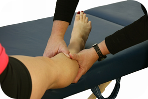 Any type of restriction that forms in the dorsi flexors, or other related areas should be removed for a full resolution of shin splints. Some of these restrictions can be removed through the process of self-myofascial release (foam rollers, and stretching). If the restrictions are severe, a manual therapy practitioner (ART, Graston, Fascial Manipulation, Massage) will be needed to break the restrictions.
- Tibialis Anterior Muscle (TA)
- The TA dorsiflexes and inverts the foot. In running, the TA is twice as active as many of the other muscles in the lower extremity. Consequently it is easily fatigued if weak. Once fatigue sets in, abnormal pronation is likely to increase.
- Extensor Hallucis Longus Muscle (EHL)
- The EHL dorsiflexes and assists with foot inversion.
- Extensor Digitorum Longus Muscle (EDL)
- The EDL dorsiflexes the foot.
Some of quietest elite runners you will see (actually not hear) are the east Africans. Many of these runners have extremely strong dorsi flexor muscles from running barefoot throughout their lives. Having strong dorsi flexors may be one of the factors as to why east Africans have dominated major marathons around the world for decades. When I ran the Paris Marathon in 2009, the winner was Tadesse Tola from Ethiopia in a time of 2 hours, 6 minutes and 40 seconds. Seeing runners maintaining such incredible speeds while quietly taking each stride is incredible.
A runner at this level recycles about half their energy through elastic recoil. This process is very similar to a spring mechanism, loading and releasing the spring with each stride. Part of this amazing spring mechanism is the dorsi flexor muscles.
Treatment of Shin Splints (MTSS)
The classic treatment of shins splints involves: rest, icing, elevation, compression and some easy stretches. This is good advice especially in the acute stage of the injury. However, exclusively following this advice will not prevent the return of the problem.
In order to resolve MTSS, the removal of any myofasial restrictions that may have formed in the soft tissue is required. This will be done in combination with a program of functional strengthening exercises for both the dorsi flexors and other areas that are affecting gait stability.
Any time a restriction is removed from one muscle the antagonistic and synergist muscles must also be assessed for restrictions. This is a key point that many manual therapists fail to recognize. For a full resolution, myofascial adhesions must be removed from the entire kinetic chain.
Exercise
 As with all musculoskeletal conditions, exercise is essential for a full resolution. Just because passive therapy has eliminated the symptoms does not mean the condition will not return. The following exercises are example of exercises that we prescribe to our patients with shin splints.
Strengthening Exercise – Example
Shin Raises – This is a great exercise for strengthening the dorsi flexors. It is best not to overdue this exercise at first, give your dorsi flexors time to adapt to the exercise. Slowly increase to the recommended number of repetitions and sets.
Version Two: This exercise is similar to version one except in two important aspects. First, the repetitions are performed at a much faster rate. Second the range of motion in which the exercise is performed is reduced.
Stretching Exercises – Example
Dynamic Shin Stretch – This dynamic stretches should be performed after a short warm-up.They are great for developing shin strength and overall motion control.
Heel Walking: There are numerous strengthening and dynamic stretching exercises that we recommend to our patients for the prevention and treatment of shin splints. The exact exercises that we recommend will vary depending on which areas of the patient’s kinetic chain are weak or restricted. In prescribing exercises, it is very common to prescribe both core and hip exercises as well as the exercises that directly affect the muscles of the shins.
If you would like more information or to purchase our books please go to www.releaseyourbody.com.
If you would like information about our clinic in Calgary Alberta please go to www.kinetichealth.ca.
(COPYRIGHT KINETIC HEALTH 2012 – ALL RIGHTS RESERVED)
]]>The following is a list of common running injures that we treat at Kinetic Health in Calgary. Under each title there are links to pages with more detailed information that we have written on these subjects. Fortunately, most of these injuries can be treated extremely well without the need for invasive procedures.
Iliotibial Band Syndrome (ITBS)
Iliotibial Band Syndrome (ITBS) is a common injury that affects runners and cyclists. Using conventional treatments, this condition often never completely resolves since they typically do not address all of the key structures involved in the injury. Click here for more information.
Resolving Hamstring Injuries
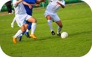
Hamstring injuries are common problems that affect a large number of athletes. These injuries can be slow to heal with a very high rate of re-occurrence. Hamstring injuries are often associated with sports that require fast acceleration and deceleration such as running (intervals), football, soccer, and rugby. Click here for more information.
 Achilles Tendonitis or Tendinopathy
The Achilles Tendon is the strongest and largest tendon in the body. It is extremely vulnerable to injury due to its limited blood supply and the numerous forces to which it is subjected. The Achilles Tendon is known as a co-joined tendon. This tendon joins directly into the calf muscles (gastrocnemius and soleus). The Achilles Tendon transmits the force generated by the calf muscles to produce the push-off required for walking, running, and jumping. Click here for more information.
Plantar Fasciitis
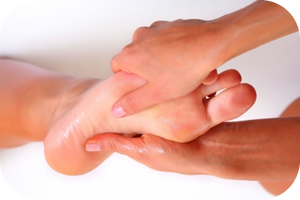
The plantar fascia is a thin sheet of fibrous connective tissue that runs from the base of your heel bone (calcaneus) and fans out to the base of your toes (proximal phalanges). The fiber orientation of the plantar fascia is very specific and very important. The plantar fascia is designed to support normal foot motions through the actions of pronation and supination. Click here for more information.
Shin Splints – Symptoms, Causes, Diagnosis, Treatment
One of the most common running injuries we treat in our clinic is shin splints or Medial Tibial Stress Syndrome (MTSS). Shin splints cause one in five athletes to stop running. In addition to running, engaging in soccer, rugby, basketball, volleyball, or any sport that involves running or jumping can cause shin splints. Click here for more information.
Stress Fractures
Stress fractures are one of the most common, and potentially serious, overuse injuries. A stress fracture is an incomplete fracture that can occur anywhere in the body, and are typically caused by repetitive forceful actions. In contrast, most other types of fractures are caused by a single, direct, traumatic impact. Click here for more information.
If you would like more information or to purchase our books please go to www.releaseyourbody.com .
Click here to book an appointment at Kinetic Health in Calgary, Alberta.
(COPYRIGHT KINETIC HEALTH 2012 – ALL RIGHTS RESERVED)
]]> exercise (Kinetic Health Calgary). Under each title there are links to pages with more information that we have written on each of these subjects. Most of these injuries can be treated effectively (90% of the time) without the need for invasive procedures.
Resolving Carpal Tunnel Syndrome With Active Release
CTS can be caused by any repetitive motion that stresses the upper extremities of the body. The increased use of computers and their accompanying flat, light-touch keyboards that allow for high- speed typing, have resulted in an epidemic of injuries to the hands, arms, shoulders, and neck. The increased use of pointing devices like the computer mouse and trackball, which require repeated subtle movements, add to these injuries. For more information click here.
 Treating Golfer’s and Tennis Elbow with Active Release
Golfer’s Elbow (Medial Epicondylitis) refers to the pain and inflammation that occurs at the inside point of the elbow (medial epicondylitis). Golfer’s Elbow can be caused by any activity (not just golf) that requires forceful and repeated bending of the wrist and fingers. For example: when the golfer swings his club, the flexor muscles and tendons of the arm tighten just before the club makes contact with the ball. For more information click here.
Shoulder Injuries – The Rotator Cuff
The shoulder joint (glenohumeral joint) is a ball-and-socket joint which joins the upper body to the arm. The shoulder joint is made up of three osseous structures, and several soft-tissue structures. For more information click here.
Resolving Sciatica with Active Release
Sciatica is a nerve compression syndrome that can be extremely painful and difficult to manage for both the patient and practitioner. Sciatic pain often affects the lower back, gluteal region, and various areas of the leg and foot. Often, the symptoms effect only on one side of the body. For more information click here.
Iliotibial Band Syndrome (ITBS)
Iliotibial Band Syndrome (ITBS) is a common injury that affects runners and cyclists. Using conventional treatments, this condition often never completely resolves since they typically do not address all of the key structures involved in the injury
.
Symptomatically ITBS presents as: A sharp or burning pain on the lateral aspect of the knee. For more information click here.
Treating and Prev
enting Meniscus Injuries
The word meniscus is derived from the Greek word that means “Crescent” as in a crescent-shaped moon. The menisci in your knee are crescent-shaped fibro-cartilaginous structures that provides stability, shock absorption, nutrition, and joint lubrication while acting to distribute your weight across your knee joint. For more information click here.
 Plantar Fasciitis
Effective resolution of Plantar Fasciitis requires a thorough understanding of the foot, how it functions, and its kinetic chain relationships to the other structures of the legs, knees, hips, and even to the core. Any successful treatment protocol must take all these elements into account, and combine treatment with an exercise routine that addresses all affected elements of the kinetic chain. For more information click here.
Resolving Whiplash Injuries with Active Release
Whiplash injuries are a very interesting, but frustrating, subject to talk about. There have been more than 10,000 research articles written about this subject. Yet, insurance companies, lawyers, and some physicians still question the validity of this syndrome. I have spent enough time in court as an expert witness to realize that opinions often have more to do with who will have to pay out money, than with the importance of addressing a condition that often leads to years of chronic pain. Pain, which in my opinion, could have been addressed with an understanding of basic biomechanics, the right treatment methodology, and exercise. For more information click here.
If you would like more information about Active Release Techniques, please check out our international best seller “Release Your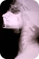 Pain – Resolving Soft Tissue Injuries with Active Release Techniques.” If you would like to more information or to purchase our books please go to www.releaseyourbody.com .
Click here to book an appointment at Kinetic Health in Calgary, Alberta.
(COPYRIGHT KINETIC HEALTH 2013 – ALL RIGHTS RESERVED)
]]>
 Cuboid syndrome is a condition that causes lateral foot pain. In forty percent of cases Cuboid syndrome is associated with lateral ankle sprains (inversion sprain). This syndrome affects the joint (capsule), ligaments, and tendons (peroneus longus tendon).
This syndrome is defined as a “minor disruption or subluxation of the structural congruity of the calcaneocuboid portion of the midtarsal joint”. In laymen’s terms, the cuboid bone has moved from its normal position in the joint.
It is a common syndrome, but not well-recognized by practitioners. Cuboid syndrome also goes by several other names: subluxated cuboid, dropped cuboid, cuboid fault syndrome, and lateral plantar neuritis.
Anatomy/Biomechanics
The calcaneocuboid joint is a vital link in lateral foot stability. This joint is susceptible to sudden injury or chronic strain, which can cause this joint to partially dislocate or subluxate.
The cuboid bone is one of the seven tarsal bones of the foot.
Joint Anatomy: The cuboid articulates with the fourth and fifth metatarsals forming a joint (tarsometatarsal joint). It also articulates with the heel bone (calcaneus), forming a joint (calcaneocuboid joint). On the inside (medially) the cuboid articulates with two bones (lateral cuneiform and the navicular). An alteration of the cuboid can have a considerable effect on the joint biomechanics of the foot.
If we were to view the bottom of the cuboid bone we would see a groove where the tendon of the peroneus longus muscle runs along it.
Peroneus Anatomy: The peroneus longus muscle originates on the lateral lower leg (upper one-third of the fibula). It then runs down around the outside of the ankle (lateral malleolus) then through the grove on the bottom of the cuboid. It then inserts into the base of the first metatarsal (and cuneiform).
The peroneus longus acts as a stabilizer of the forefoot. The cuboid bone acts as a pulley which increases mechanical advantage for the peroneus longus muscle.
 Causes
A single impact injury or repetitive motion can cause this syndrome to occur. Runners and dancers (especially ballet dancers) are susceptible to this injury because of the high levels of repetitive impact. In addition basketball players or racquet sport players also are susceptible due to the lateral motions required to play these sports.
If a person abnormally pronates their foot during the push off phase of gait, they will be more susceptible to a cuboid injury. Excessive pronation causes an increase in force transferred to the cuboid bone which causes instability and a resultant injury.
Symptoms
Patients who have cuboid syndrome often complain of lateral foot pain, or weakness in their feet. Pain from cuboid syndrome can also radiate to the front of the ankle. This pain is often more noticeable during time of exertion (toe-off portion of the gait cycle), or on impact.
The type of pain found in cuboid syndrome may not be a very good indication of this condition.Pain can be intermittent, or persistent, it can also develop suddenly or slowly over a period of time.
 Diagnosis and Imaging
Upon physical examination, the patient may have pain directly over the cuboid bone (especially when pressure is applied dorsally on the plantar surface). In some cases there may be bruising, redness and swelling. Range of motion in the ankle is often limited in cuboid syndrome (dorsi and plantar flexion).
X-rays, CT scans or MRIs are of little value in the diagnosis of cuboid syndrome. The only reason an X-ray is of value in the diagnosis of this syndrome is to rule out fractures or some type of pathological condition.
Treatment
Several forms of manual therapy can be used in treating this condition. The sooner that treatment is implemented the faster the results will be.
Manipulation: One of the most successful treatments we have found is manual manipulation. A therapist training in extremity manipulation (chiropractor, physiotherapist, podiatrist) can often reduce the pain of cuboid syndrome in a short period of time.
Soft tissue techniques: Techniques such as Active Release, Graston, Massage Therapy, and Fascial Manipulation can also be of great help. The key is to work with all of the structures involved in the kinetic chain, not only at the site of pain. These structures could be anywhere from the foot right up to the hip.
- For example we know that the peroneus longus muscle is involved in plantar flexion of ankle, and that this greatly affects cuboid stability. A problem with the peroneus longus could create an alteration in the normal plantar flexion movement. Even though the peroneus longus is most likely involved in this injury, we cannot take it for granted that this is the only plantar flexor affected. Treatment of all plantar flexors and their antagonist muscles, the dorsi flexors, is often needed for a full resolution.
- Plantar flexors: Calf muscles (Gastrocnemius & Soleus), Tibialis Posterior, Flexor Digitorum Longus, Flexor Hallucis Longus, Peroneus Longus, Peroneus Brevis, and Plantaris.
- Dorsi flexors: Tibialis Anterior, Extensor Digitorum Longus, Extensor Hallucis Longus.
 Exercise: Exercises should focus on stretching the peroneus longus and the calf muscles. These stretches are combined with strengthening the extrinsic muscles of the foot. In addition, it is essential to perform balance exercises so that the injury does not reoccur.Please go to www.releaseyourbody.com for a specific exercise needed to rehabilitate this injury.
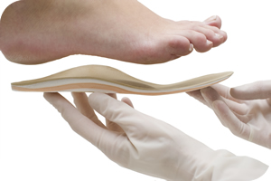
Orthotics: Custom fit orthotics will help to correct abnormal pronation and supinationthat may be leading to cuboid syndrome.
If you would like more information or to purchase our books please go to www.releaseyourbody.com .
If you would like information about our clinic in Calgary Alberta please go to www.kinetichealth.ca.
(COPYRIGHT KINETIC HEALTH 2012 – ALL RIGHTS RESERVED)
]]> Approximately 90 percent of headaches originate as tension headaches. Medical experts continue to debate over the causes of tension headaches. In my opinion, 90% of tension headaches are either caused by mechanical factors or are perpetuated by them. In other words, most ongoing, chronic, tension headaches can be attributed to specific physical restrictions. These are restrictions in muscles, ligaments, tendons, and fascia which cause nerve impingement syndromes, vascular changes, motion compensations and the output of biochemical substances that affect pain centres.
Symptoms of Tension Headaches
Tension headaches usually last from half-an-hour to an hour but they can continue to return for weeks, and in chronic cases, last for years. People who suffer from tension headaches often describe their headache as a dull ache, or they may experience them as a band of tightness around the sides of their head. This band may even feel like a vice compressing their skull. In severe cases the pain may even feel like a hooded cape that drapes over and across the shoulders. Obviously in such severe cases more than just the head is involved in creating the pain syndrome.
The following is a list of some of the common symptoms of tension headaches:
- Band like pressure around the head.
- Difficulty concentrating.
- Difficulty sleeping (insomnia).
- Fatigue and irritability.
- Loss of appetite.
- Neck, jaw/TMJ, or shoulder discomfort.
- Severe pain behind the eyes.
- Tenderness of the scalp.
You can usually differentiate a tension headache from other types of headaches because there are many symptoms you will not experience. For example, tension headaches do not cause visual disturbances, nausea, vomiting, numbness on one side the body, or slurred speech.
Triggers for Tension headaches
Tension headaches are triggered by numerous causes. Some of the potential triggers include: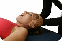
- Anxiety (including several medications used for reducing anxiety).
- Arthritis
- Clenching or grinding teeth (bruxism).
- Dental Work.
- Depression (including medications used for depression).
- Holding one position for a long time, or working in awkward positions.
- Inflammation of the neck or shoulders.
- Lack of sleep or insomnia.
- Lack of physical activity.
- Overuse of headache medication (3rd most common cause of headaches).
- Poor posture.
- Stress.
- Trauma.
- Whiplash injuries (hyper-extension hyper-flexion injury)
 Nerve Compression
Nerve compression can be a major factor in the initiation and perpetuation of tension headaches. If we look at the pathway of different peripheral nerves that transect, or pass under, musculoskeletal structures (muscles, ligaments, tendons, and fascia) it is easy to see how nerve compression can create a tension headache. Consider how a restriction of the following muscles can cause nerve compression which in turn can cause a tension headache.
Muscles:
- Superior obliques
- The Suboccipital nerve supplies input to the muscles of the suboccipital triangle. Compression of this nerve can occur at a muscle called the superior oblique.
- A trigger point in the superior oblique muscle itself will also refer pain to various regions of the head.
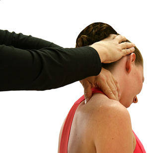
- Semispinalis (capitis, cervicis) muscle
- The greater occipital nerve is located directly under the semispinalis capitis muscle. Compression of this nerve is one of the causes of cervicogenic headaches. The symptoms from these headaches are called occipital neuralgias.
- Occipital neuralgia is a medical condition characterized by chronic pain in the upper neck, back of the head and behind the eyes. This is sometimes known as C2 neuralgia or Arnold’s neuralgia.
- SCM (Sternocleidomastoid) muscle
- The lesser occipital nerve and the greater auricular nerve both pass by theSCM. Compression of these nerves will give symptoms of occipital neuralgia.
- Trapezius (upper fibers) muscle
- The third occipital nerve travels under the trapezius muscle until it pierces this muscle and ends up in the lower part of the head (occiput). Compression of this nerve by a restriction in the trapezius muscle can also cause occipital neuralgias.
Biochemical Changes
 If we review the literature on tension headaches there is no doubt that tension headaches are associated with changes in the levels of certain brain chemicals such as serotonin, bradykinins, and substance P.
Serotonin acts as a chemical messenger that transmits nerve signals between nerve cells. It also causes blood vessels to narrow.
Bradykinins mediates the inflammatory response, increases vasodilatation(expansion of arteries and veins), and causes contraction of smooth muscle.
Substance P functions as a neurotransmitter, especially in the transmission of pain impulses from peripheral receptors to the central nervous system.
The question is, are these chemical the initial factor that created the headache or are they a secondary consequence of physical restrictions. In my opinion, the physical changes probably took place first; which in turn caused a cascade of events that eventually increased, or altered, the levels of these chemicals.
Support for my perspective comes from what is called central sensitization. Central sensitizationhas been hypothesized as one of the primary mechanisms of chronic pain conditions. Central sensitization occurs when pain receptors in your nervous system (nociceptive neurons) become sensitized by tissue damage or inflammation.
By damage, I am not just referring to some type of trauma like a motor vehicle accident. I am also referring to damage caused by:
- The micro trauma of repetitive strain injuries.
- Stress caused by muscle imbalances.
- Long-term stress placed on your body by poor posture.
- De-conditioned muscles.
- A thickening of the fascia. It is postulated that this thickening is caused by an increase in hyaluronic acid, which causes ‘densification’ of the fascia.
All these causes can produce areas of hyper-tonicity (increased tension) in our bodies that become chronically restricted; such as that huge knot most people have in their shoulders or the one just under the base of their skull.
When these hypertonic tissues becomes irritated or inflamed it releases chemical mediators (bradykinin, serotonin, substance P), which cause a sensitization of nerve endings resulting in pain, tension, and the production of headaches. If we review the history of most patients who suffer from tension headache we will routinely find a source of initial tissue damage (accident, trauma, history of tension) and associated inflammation which causes biochemical changes that elicit headaches.
Treatment of Tension Headaches
 To resolve Tension Headaches it is essential that we remove all physical restrictions throughout the body’s Kinetic Chain. This would include restrictions in your shoulders, neck, jaw, skull, and perhaps other affected areas. In other words, wherever we find restrictions that may be initiating a physical cascade of events that will eventually cause physical or biochemical changes resulting in a headache. This will include:
- Soft tissue adhesions – These will be found in muscles, ligaments, tendons and fascia. Anywhere that a change in tissue texture, tension, movement or function is noted.
- Peripheral nerve entrapments – Besides the actual tension headache, nerve entrapment symptoms include: paresthesisas or abnormal neurological sensations which include:numbness, tingling, burning, prickling, hyperesthesia (increased sensitivity) and muscle atrophy.
It is extremely important to recognize that if we are only dealing with the headache on a pharmaceutical basis we have not addressed the chronic chain of events that perpetuates the headache.
There is a considerable amount of research in the literature to support this perspective. Even a change in posture can greatly affect the prevalence of a tension headache.
Research has demonstrated that tension or weakness in any of the following areas will cause an increase in both the intensity and frequency of tension headaches.
Examples of Anterior Structures:
- Neck flexor muscles
- Sternocleidomastoid muscle
- Temporalis muscle (above Jaw)
- Examples of Posterior Structures:
- Suboccipital muscles
- Splenius Capitus
- Semispinalis Capitus
- Trapezius
Our clinical experience has shown that the majority of chronic tension headaches can be completely resolved or substantially reduced by using soft tissue therapy (Active Release Techniques, Graston Techniuqe, Fascial Manipulation, or Massage therapy) in conjunction with the correct exercise program.
This information is derived from our Release Your Kinetic Chain Exercise Series. If you would like to more information or to purchase our books please go to www.releaseyourbody.com .
If you would like information about our clinic in Calgary Alberta please go to www.kinetichealth.ca.
Referred pain from the trochlear region in tension-type headache: a myofascial trigger point from the superior oblique muscle. Headache 2005 Jun;45(6):731-7. Fernandez de las Peñas C, Cuadrado ML, Gerwin RD,Pareja JA.
Myofascial trigger points and sensitization: an updated pain model for tension-type headache Fernandez-de-las-Penas C, Cuadrado ML, Arendt-Nielsen L, Simons DG, Pareja JA. Cephalalgia. 2007 May;27(5):383-93. Epub 2007 Mar 14. inDepartment of Physical Therapy, Occupational Therapy, Physical Medicine and Rehabilitation, Universidad Rey Juan Carlos, Madrid, Spain.
Stress-induced pain and muscle activity in patients with migraine and tension-type headache. Leistad RB, Sand T, Westgaard RH, Nilsen KB, Stovner LJ. Cephalalgia. 2006 Jan;26(1):64-73. Department of Neuroscience, Norwegian University of Technology and Science and Department of Neurology and Clinical Neurophysiology, St Olavs Hospital, Trondheim, Norway.
(COPYRIGHT KINETIC HEALTH 2012 – ALL RIGHTS RESERVED)
]]>
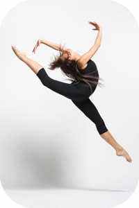 An injury to the Flexor Hallucis Longus (FHL) tendon causes medial ankle pain or pain on the bottom of the foot. FHL injury is a condition that is often overlooked or misdiagnosed. This injury affects dancers, runners, soccer players, and any other athlete who performs repeated, propulsive forces, or jumping. Injury of the Flexor Halicus Longus muscle is sometimes called “Dancer’s tendonitis” but it is not limited to just dancers.
The Flexor Hallucis Longus (FHL) muscle allows you to point your big toe (plantar-flexing your big toe) and stabilizes the Subtaler Joint. The Subtaler Joint is located between two bones in your ankle – the Talusand the Calcaneus. The Subtaler Joint allows movement of the heel toward the medial plane (inversion) as well as movement of the heel towards the lateral plane (eversion).
Anatomy/Biomechanics of Medial Ankle Pain
The Flexor Hallucis Longus muscle is located deep under your calf muscles (the most lateral muscle of the deep compartment). The FHL originates on the lateral lower leg (distal 2/3 of Fibula). It then travels at an oblique angle (crosses the posterior surface of Tibia) down towards the medial ankle (posterior surface of Talus) and travels under a section of the heel bone (Sustentaculum Tali of Calcaneus). The FHL then passes under the sole of the foot (between the two heads of the FHB) and inserts into the base of the big toe (base of the Distal Phalanx of Hallax).
As mentioned the FHL Tendon curves around a structure called the Sustentaculum Tali. TheSustentaculum Tali is a bony section on the inside of the heel bone (Calcaneus). This is often a site of friction and irritation for the FHL tendon. The FHL tendon also travels between the twoSesamoid bones just behind the big toe (Metatarso-Phalangeal Joint). The sheath that surrounds the FHL tendon is often irritated.
Causes and Presentation of Medial Ankle Pain
The repetitive motion of pushing-off with your foot (plantar flexion) during dance, running, soccer, and jumping can cause injury to the FHL.
Injury to the FHL tendon and muscle can present in a variety of ways – sometimes involving inflammation and sometimes not. Injury to the tendon without inflammation is refer to as a “Tendinopathy”. Tendinopathy refers to a degenerative lesion in the tendon without affecting the tendon sheath that surrounds the tendon.
When FHL inflammation (tendonitis) is present in the foot, it usually occurs in one of the following three locations.
- Inside of the ankle (fibro-osseous tunnel along the Posteromedial Ankle).
- At the “Knot of Henry” – a section just behind the big toe (first metatarsal) where the FDL muscle crosses the FHL tendon.
- Just behind the big toe by the Sesamoids bone.
o A Sesamoid bone holds its tendon away from the center of the joint and acts to increase mechanical force.
When the FHL tendon becomes nodular, a condition called Hallux Saltans can develop. Hallux Saltans is similar to trigger finger in the hand, except it occurs in the big toe. Triggering of the toe occurs when the nodular thickening of the tendon snaps through the fibro-osseous tunnel. This causes a jerking motion, much like a trigger finger.
If not addressed Hallux Saltans can contribute to the progression of an additional condition called Hallux Rigidus. Hallux Rigidus means “stiff great toe”. Hallux Rigidus is the second most common disorder of the first MTP joint. The most common injury is a bunion – otherwise known as Hallux Valgus.
Special Considerations For The Dancer
 Injury to the FHL in dancers is often caused by the repetitive motion of changing position from a plié position to a relevé position. This action produces a force that is 10 times the dancer’s body weight.
Ideally, for greater stability and increased propulsion, the foot should be in a supinated position at the heel during push-off. For a dancer, any action that causes a reduction in plantar flexionmotion can create an FHL injury. Biomechanically, a lack of plantar flexion leads to a prolonged pronation position of the foot when pushing off in the Propulsion Phase.
In dancers, the FHL tendon is often compressed while performing a relevé position and is over-stretched while performing a plié postion. In such a case, the dancer will feel posterior medial ankle pain when performing the plié.
Diagnosis and Imaging – FHL Injury
X-rays for this condition are not very specific. X-Rays are good for ruling out fractures (Calcaneus, Distal Medial Malleolus, or Os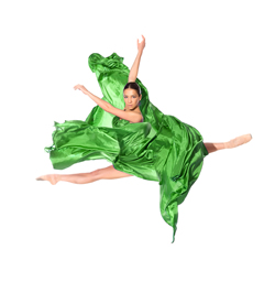 Trigonum) that may cause an impingement of the FHL tendon. It is important to note that X-Rays will not provide a definitive diagnosis of FHL injuries.
A good history is much more definitive, especially when accompanied by a full physical examination and an MRI. MRI imaging is an excellent resource for showing damage to tissue fibers, inflammation, and swelling (edema).
If the MRI is not available or is too expensive, a CT scan (Computed Tomography) can prove to be a useful alternative.
Treatment
 In most cases, FHL syndrome (involving partial tears, tendinosis, or inflammatory conditions) responds well to conservative care. However, a complete tear of the FHL tendon may require corrective surgery since serious cases of FHL injury could end an athlete’s career.
Conservative care includes:
- Ice or heat depending on stage and type of injury (inflammation or not). See our Icing andHeating blog.
- Reduction in activity. Usually, some modification in all activities is necessary.
- Supports (crutches or walker boot) can be beneficial.
- Taping – Kinesio Taping. At Kinetic Health we use SpiderTech Kineso-taping.
- Soft-tissue and joint mobilization (Active Release, Graston Techniques, Massage, and Fascial Manipulation).
- Inflammatory strategy implementation (medication for no longer than seven days). See ourblog about Reducing Inflammation.
- Exercises for increased strength, flexibility and balance. Go to www.releaseyourbody.comfor exercises you can use to treat this condition.
- Biomechanical corrections (footwear or Orthotics) .
- A gradual return to activities.
If you would like more information or to purchase our books please go to www.releaseyourbody.com .
If you would like information about our clinic in Calgary Alberta please go to www.kinetichealth.ca.
(COPYRIGHT KINETIC HEALTH 2013– ALL RIGHTS RESERVED)
]]>
 Think long-term…. avoid the rebound effect!
Keep your weight-loss goals realistic. It is reasonable to expect to lose about one to three pounds per week. Too rapid weight-loss almost always results in a rebound effect, where all the lost weight returns if you fall off the path. In many weight-loss programs, the quick weight that you lose is often due to loss of muscle, rather than fat loss. When you lose muscle mass, you actually lower your metabolism since muscles are the primary “burners” of carbohydrates and fats in your body.
Dramatic decreases in caloric intake leads to decreased or slower metabolism, and more weight gain.
Suddenly reduced, or too low, caloric intake leads to a loss of muscle mass as your body struggles to meet its daily energy requirements by burning muscle rather than fat. It also causes your body to STORE FAT. This can happen whenever you eat less than 1000 calories a day. Your body requires at least 1500 calories a day just to perform its basic functions. Decreases in your muscle mass results in a decrease in your metabolic rate, which in turn results in more fat being stored! This is the major reason for the failure of over 80% of all diet plans. Instead of training your body to burn fat, they train your body to store fat.
Graze, Don’t Gorge!
Frequent SMALL meals are better for weight loss. Frequent small meals serve to convince your body that you are NOT starving, that food is abundant, and that fats DON’T need to be stored for hard times. Basically, you need to provide you body with good quality food on a regular basis. This will have several positive effects: it will help balance your blood sugar, accelerate healing, and provide you with the energy you need for exercising.
Your Mother was right…. you need to eat breakfast!
Did you know that your body burns about 450 calories when you sleep at night? Your body needs to re-fuel at breakfast time. If you don’t have breakfast, your blood sugar levels can fall too low, resulting in cravings for high-sugar food. Successful individuals in any weight-loss program always eat breakfast.
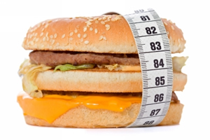 Forget the fast food.
Fast food is generally made with cheaper ingredients, such as high fat meat, lots of saturated and trans fats, lots of salt, and cholesterol. All of these ingredients lead to health problems, weight gain, high blood pressure, inflammation, and heart disease. Consider what is in a basic, medium-size, fast food meal of a burger, French Fries, and a vanilla shake. In that one meal you will get 87 grams of fat and 1,480 calories. If you want to gain weight, inflame your body, and die of a heart attack, then by all means, go for it and enjoy that meal.
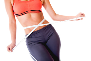
Start thinking about your body-fat percentage not your weight.
Body-fat percentage measures the percentage of fat in your body. If you are 200 pounds and 30% fat, it means that your body consists of 60 pounds of fat and 140 pounds lean body mass. Your goal is to gain muscle (which burns fat). Remember, muscle also weighs more than fat. So initially, don’t worry about what the scales say. Your focus is to get rid of the fat while adding on muscle.
Add Muscle to Your Body
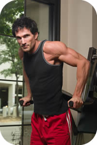 Muscles burn more calories than any other type of tissue in your body (with the exception of your brain). Muscles help to increase your resting metabolic rate, which means you burn fat even when resting. For each pound of muscle that you add to your body, you will burn an extra 50 calories each day. This means if you add five pounds of muscle mass to your body, you can burn 250 extra calories a day, even when you are just sitting around.
Strength training (or muscle building) should be one of the fundamentals of any weight loss program. (Note: diet changes alone will not give you the results you want!) Strength training builds muscle mass and increases your resting metabolic rate. Exercise and strength training are the keys for getting rid of all that already stored, excess fat.
It is important to differentiate between weight-lifting exercise and cardiovascular exercise. There is no doubt, that cardiovascular exercise does burn a lot of calories. The problem is, it is not nearly as efficient at turning up your fat burning furnace as weight lifting can be. In fact when you compare the two exercise formats, cardiovascular exercise is about 40% less efficient at burning fat than weight lifting . I am not saying you should give up your cardio-routine. Cardiovascular exercise is great for your overall health, especially your heart. The problem is that using only cardio as your primary exercise will not help you to achieve the fat loss you what.
I am currently developing an weight-loss exercise routine that is designed to help you increase muscle mass (lose fat). I will be presenting this in a future blog.
Get a good night’s sleep!
You need to sleep at least 8 hours every night if you want to lose weight. Lack of sleep increases stress, stress increases cortisol levels in your body, and increased cortisol levels result in weight gain. Lack of sleep affects the level of hormones which control weight-loss – melatonin and human growth hormone. Both of these are produced during sleep, and interruptions and lack of production inhibits their positive effects on your body. In fact, research has shown that the probability of your being obese is 73% higher if you get only four hours of sleep per night.
Specific Dietary Recommendations
If you want to lose fat, decrease your body fat percentage, and feel the best you have felt for years here is what I suggest.
 Stay hydrated:
Drink two glasses of water before each meal. Then continue to drink water on a regular basis throughout the day. Read my blog about the importance of hydration for more information.
Drinking lemon or lime water is a great way to make your body more alkaline. The amazing thing is that lemons can turn even regular tap water alkaline.
Meals and snacks:
My basic dietary program is quite simple, eat three meals per day and 2 or 3 snacks. Avoid processed or packaged foods, refined carbohydrates, high fat foods, and eat your foods in as natural a state as possible. You should also do everything can to make your food taste great. Use spices and herbs liberally but take it easy on the salt you add to your food.
Each meal should contain protein, complex carbohydrates and essential fatty acids. You should also try to make choices that will make you meals more alkaline in nature.
Remember,eating health foods can be a very enjoyable experience. You can lose all the fat you want, and still enjoy your food.
Breakfast
First off you need to eat breakfast, DO NOT SKIP THIS MEAL! Breakfast should contain a serving of protein, dairy, fruit or grains and any amount of vegetables you would like.
You can have coffee (espresso is more pH balanced – drip coffee is quite acidic), tea (ginger or green tea), or “FRESH” vegetable or fruit juice with your breakfast.
Please note: If you are having grains, then make sure you are not reacting to wheat or gluten. Again a topic for a future blog.
Lunch
At lunch, you should have a serving of protein, fruit, and any amount of vegetable that you would like. Remember to drink two glasses of water (lemon water preferably) before you eat your lunch.
 Dinner
Again, as with the other two meals, you need to get a serving of protein, complex carbohydrates and essential fatty acids.
For many individuals this is the main meal of the day. Try not to bulk up on this meal, and be sure to spread your total food intake throughout the day. As with the other meals, take a look at the PRAL chart to balance out the acidity of the meal.
Snacks:
There are numerous healthy foods that you can use as snack throughout the day. Just be sure not to eat too much of these foods, otherwise you may find that you are sabotaging your fat-loss program.
I hope you will consider following the fat loss program I have just laid out for you. This program can literally transform your life. It has taken many of my patients out of pain, returned their energy, and literally revitalized their lives. I hope it can do the same for you.
More Information
See the following blogs for the rest of Dr. Abelson’s Essential Nutritional Program:
- Dr. Abelson’s Essential Nutritional Program Part 1 – Eating the Right Foods
- Dr. Abelson’s Essential Nutritional Program Part 2 – All About Fats – Good and Bad
- Dr. Abelson’s Essential Nutritional Program Part 3 – The Importance of Drinking Water
- Dr. Abelson’s Essential Nutritional Program Part 4 – Alkaline Diets
- Dr. Abelson’s Essential Nutritional Program Part 5- Weight Loss and Conditions Related to Obesity
Do you want more information about this, and other topics. Our books (eBooks and hard-copy) provide more information about soft-tissue injuries, rehabilitative exercises for injury recovery, and how to use these to activate and restore all the structures of your kinetic chain.
To purchase our internationally best-selling books, visit www.releaseyourbody.com.
For information about our clinic in Calgary, Alberta, please go to www.kinetichealth.ca. Or call us at 403-241-3772.
(COPYRIGHT KINETIC HEALTH 2013 – ALL RIGHTS RESERVED)
]]> Is this section of the my dietary protocol, I will cover some of the consequences of obesity. My hope is that you will take this issue seriously, not because of the superficial way society judges overweight individuals, but because of how it is affecting your health – physically, emotionally and mentally.
Then, on the next installment of this blog, I will provide you with viable solutions that I have used with my patients to bring their body fat percentages into a healthy range.
Please note that weight loss is now a multi-billion dollar industry that is full of hype, empty promises, and quick fixes that seldom deliver on their promises. My objective of this weight-loss section is to help you cut through that hype so you can find effective ways of dealing with this common condition.
After we get through the section on conditions related to obesity, I will give you viable solutions to help deal with this issue. As we go through this information please keep in mind two key factors:
- Just because you have never been able to reach your weight loss goals does not mean there are no viable solutions for you. Over 90% of individuals who follow my weight-loss recommendations reach their goals.
- Please, don’t beat yourself up over your current weight. Our society has some very warped ideas about what your ideal weight should be. As I say to my wife, I would be very disappointed if she lost all her curves.
Before we get into specific recommendations about how to get your body-fat percentage within normal ranges, let us first review why we should take the extra weight we are carrying around seriously.
Conditions Related to Obesity
It always amazes me that patients do not see the correlation between their current health status, and the extra weight that they are carrying around in their bodies.
 Here is a list of conditions/states that are directly related to obesity:
- Asthma.
- Benign Prostatic Hyperplasia.
- Cancer (obesity has been linked to the following types of cancer: breast, colon,endometrium, kidney, esophagus, gallbladder, pancreas , and ovarian).
- Cardiovascular disease.
- Cognitive decline (Individuals who are obese, with high blood pressure, and other risk factors (metabolic abnormalities) can experience a faster decline in their cognitive function. August 21, 2012, Neurology)
- Depression (Research has shown that obese people have clinical depression scores that are as bad, or worse than, for patients suffering from chronic pain).
- Diabetes.
- Erectile dysfunction (obesity damages your cardiovascular system and decreases your testosterone levels).
- GERD.
- Gout.
- Heart disease (coronary heart disease, congestive cardiac failure).
- Headaches.
- High Cholesterol.
- Hormonal dysfunctions/imbalances.
- Hypertension – High blood pressure (two-thirds of obese individuals are at risk of hypertension).
- Inflammatory conditions.
- Insomnia.
- Migraine headaches.
- Musculoskeletal Injuries such as back pain, ankle pain, knee pain, hip pain, shoulder pain, neck pain, wrist pain, elbow pain, wrist pain, and hand pain.
- Osteoarthritis.
- Osteoporosis.
- PMS.
- Sexual dysfunction (impairs sexual performance and decreases sex drive).
- Slow healing.
- Strokes.
- Testosterone decrease. (In both men and women this fat-burning, muscle-building hormone is critical to weight loss).
- Urinary Incontinence.
At Kinetic Health, our clinical specialization is the treatment of musculoskeletal injuries. In most cases, we can help the patient resolve 80%-90% of their injuries in only a few short weeks (depending on severity of the condition and age). In the remaining cases, where I see little or very slow progress, the lack of response is often related to the patient’s body-fat percentage issues.
Like it or not carrying too much weight around is like a ticking time bomb, especially if that weight is around your mid section.
People who store fat around the middle (abdomen, chest, and internal organs) have an “apple” shape, and are more prone to diabetes (type II diabetes is at epidemic levels), heart disease, high blood pressure, gall bladder problems, systemic inflammation, and the wide array of health issues associated with inflammation.
People with a “pear” shape, who store weight on their hips and thighs are at a lower risk for these conditions.
Inflammation and Obesity
Here is one fact you cannot get around. If you are obese/overweight, your body produces an increased level of pro-inflammatory compounds (such as prostaglandin E2). On the surface, these inflammatory compounds may not seem like a big deal, until you do a little investigation. Take a look at some of common conditions that are related to increased levels of inflammation in your body:
- Alzheimer’s disease
- Arthritis
- Atherosclerosis
- Cancer
- Chronic pain
- Crohn’s disease
- Dementia
- Diabetes
- Emphysema
- Heart disease
- High blood pressure
- Insulin resistance
- Musculoskeletal conditions
- Osteoporosis
- Parkinson’s disease
Unfortunately, obesity and inflammation creates an escalating cycle of dysfunction. Inflammatory hormones ,such as prostaglandin E2, inhibits your body’s ability to burn fat. This lowers your metabolism, makes you gain even more weight which increases the levels of inflammatory hormones. This really is a nasty cycle of dysfunction
Osteoporosis and Obesity
 There are numerous steps you can take to slow down osteoporotic degeneration.
The first step is to get rid of any extra weight you are carrying.
Osteoporosis is a condition that occurs when the quality and quantity of bone tissue is weakened, making your bones more susceptible to bone loss and breakage. It also prevents your bones from re-modeling and re-building themselves.
The relationship between obesity and osteoporosis has been widely studied. Initially studies indicated that obesity (increase weight ) may help prevent osteoporosis, but this is not true. More recent studies have now indicated that the same nutritional problems associated with obesity lead to Osteoporosis. The two conditions are linked.
Relationship of Obesity with Osteoporosis Lan-Juan Zhao, Yong-Jun Liu, Peng-Yuan Liu, James Hamilton, Robert R. Recker and Hong-Wen Deng – The Journal of Clinical Endocrinology & Metabolism Vol. 92, No. 5 1640-1646
Osteoporosis – Some Important Facts
Osteoporosis should be take seriously, and you should do everything you can to avoid it. Consider some statistics about osteoporosis.
- Over 80% of all fractures in people over fifty years of age are caused by osteoporosis.
- Osteoporosis causes 70-90% of hip fractures annually.
- 37% of men and 28% women and who suffer a hip fracture will die within the next year.
- Only 44% of people, discharged from hospital after a hip fracture, return home.
Osteoarthritis and Obesity
Osteoarthritis is a complex degenerative condition that affects the joints in your body. Primary Osteoarthritis affects the weight-bearing joints in your knees, feet, hips, and lower back, as well as the fingers and neck.
Osteoarthritis is a condition that is caused by excessive and long-term wear-and-tear of your joints, and is often accompanied by inflammation of the area with pain, swelling, and stiffness.
Primary Osteoarthritis begins by affecting the cartilage of your joints. Healthy cartilage allows bones to glide over each other and assists in absorbing the shock and stress of movement. In osteoarthritis, the top layer of the cartilage breaks down and wears away, allowing the bones under the cartilage to rub against each other. The rubbing causes inflammation or swelling, pain, and decreased mobility of that joint. Over time, bone spurs may grow and the joint may lose its normal shape.
Obesity increases the chances of developing osteoarthritis since the excess weight multiplies the pressure and forces on the joint, leading to more wear and tear over time.
Osteoarthritis and Weight Loss – the GOOD NEWS
 Even moderate decreases (15 lbs over 6 weeks) in weight can significantly reduce the amount of pain caused by osteoarthritis. Stephen Messier, a lead author of a noted study showed that “The accumulated reduction in knee load for a 1-pound loss in weight would be more than 4,800 pounds per mile walked”. In other words, for every pound of weight-loss, you will find a four-pound reduction in the stress experienced with each step!
The effects of even a moderate weight-loss is especially noticeable in weight bearing joints such as the knee and hip. This can result in significant improvement in quality of life. This is an achievable goal, and can help you to live a more pain-free and enjoyable life.
More Information
See the following blogs for the rest of Dr. Abelson’s Essential Nutritional Program:
- Dr. Abelson’s Essential Nutritional Program Part 1 – Eating the Right Foods
- Dr. Abelson’s Essential Nutritional Program Part 2 – All About Fats – Good and Bad
- Dr. Abelson’s Essential Nutritional Program Part 3 – The Importance of Drinking Water
- Dr. Abelson’s Essential Nutritional Program Part 4 – Alkaline Diets
- Dr. Abelson’s Essential Nutritional Program Part 6
Do you want more information about this, and other topics. Our books (eBooks and hard-copy) provide more information about soft-tissue injuries, rehabilitative exercises for injury recovery, and how to use these to activate and restore all the structures of your kinetic chain.
To purchase our internationally best-selling books, visit www.releaseyourbody.com.
For information about our clinic in Calgary, Alberta, please go to www.kinetichealth.ca.
(COPYRIGHT KINETIC HEALTH 2013 – ALL RIGHTS RESERVED)
]]> Alkaline Diets
The concept of balancing your bodies pH through diet has been around for a very long time (acid-base homeostasis or acid-base balance). Thirty years ago, I was using alkaline foods, such as wheatgrass, in my diet to help me recover from workouts for Ironman races. Today I am still using the same concepts to help me recover from fitness workouts, occasional injuries, and to keep my biological clock from rushing forward (I will explain that one later).
The Alkaline Diet postulates that our bodies are designed for predominately alkaline foods, not the acidic diets of today (high meat, high grain, processed foods). There is considerable evidence to support the concept that acid-based diets are directly related to the degenerative, cardiovascular, and pathological conditions that are so prevalent today. Even inflammation, and weight gain have been associated with acidic diets.
Before we get into dietary details lets do a quick review of acidity and alkalinity.
Your Body’s pH
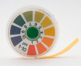
pH is a measure of the acidity or alkalinity of a solution. Solutions with a pH less than seven are acidic, while those with a pH greater than seven are basic (alkaline). A pH of 7 is neutral. The pH of your blood is maintained in very narrow range from 7.38 to 7.42. Even a minor deviation from this range can have severe consequences, sometimes even resulting in death. Your body maintains this narrow balance through three mechanisms: your respiratory system, chemical buffers, and your kidneys (excreting acid).
- Your Respiratory System: When acidity increases there will be a corresponding increase in pulmonary respiration. This will cause an increase in the excretion of H+. Hydrogen molecules are what make a solution acidic. Then a secondary reaction occurs (H + HC03 = H2CO3) forms water (H2O) and carbon dioxide (CO2), which is then eliminated through respiration.
- Chemical Buffers – When your body buffers a solution, the buffer binds to the loose hydrogen. Removal of loose hydrogen from a solution makes it less acidic. These reactions can occur within seconds.
- Your Kidneys – The kidneys can balance blood pH by excreting excess acids (or bases). This is a much slower process than the respiratory mechanisms or the chemical buffers.
The balance between acids and bases in your body is of enormous importance to the functioning of all your metabolic processes. This balance is critical for the functioning of your proteins, the permeability of your membranes, the delivery of electrolytes, and the functioning of every cell, organ, and structure in your body.
pH and Aging
Earlier, I mentioned that we can use the concept of alkaline diets to keep our biological clocks from rushing forward. Let me explain why.
As we age, the body’s ability to remove acidity decreases (by up to 80% by the later part of your life). This is primarily due to decreased kidney function that occurs with normal aging (especially after age 50). This is due to a decrease in glomerular filtration. The important point here is that your acid-base balance is dependent upon your kidney’s ability to remove acid from your body, and that this ability decreases with age.
This decreased kidney function can lead to a state known as low-grade metabolic acidosis (increase in H+). Low-grade metabolic acidosis is a clinical disorder that is characterized by an increase in total body acidity. Even mild acidosis increases bone loss, muscle wasting, and inflammation. Not what you want to do if you are trying to repair your body or increase your athletic performance. If we can avoid increased acidity in our bodies, we can slow down or avoid some of the diseases associated with aging and acidity.
Diet and pH
 The food you eat can have an effect on the how much stress is place on your body as it attempts to maintain its pH balance. Generally speaking, the North American diet is extremely acidic, because of the quantity of grains, animal products, and processed foods that is consumed. This constant acid-state does put a considerable load on your body’s buffering systems.
As we already mentioned the pH of your blood is kept within a very narrow pH range – around 7.4. Your body’s buffering systems does this by drawing upon minerals from your bones (calcium, magnesium, potassium, zinc), amino acids from your muscles (glutamine), or by using your respiratory or kidney’s buffering systems.
In contrast to the pH of your blood, the pH of your urine varies based on your diet. Most people in North America have a very acidic diets, which shows up directly in their urinary pH. Even consuming a high-salt diet can cause your body to create a state of low-grade acidosis. Something to think about – that extra table salt you are adding to your food may be leading to chronic conditions such as osteoporosis, slowing your healing, inflaming your body, and even making you gain weight.
Acidic states damage your body
As mentioned, most North American diets produce a state of “chronic low grade acidosis”. What this means is that our bodies are continuously pulling key minerals out of our bones, and amino acids from our muscles, in an attempt to maintain a constant pH in our blood and cells. Bone and muscle breakdown are two classic examples:
- Bone Degeneration – Close to a century ago, medical research concluded that constant acidic states in our bodies cause the release (leaching) of calcium from our bones. This is because calcium is a strong base, used by the body to neutralize the acid. The net result is osteoporosis, or weak, fragile, thin bones.
- Muscle Break Down – Your muscles contain high levels of an amino acid called glutamine. This amino acid helps you to build and preserve muscle mass. Your body also uses this amino acid to balance your pH. When your body becomes too acidic, it will start to break down the glutamine in your body. This drives the pH scale to a more alkaline direction.
 Side Note: Glutamine is definitely a supplement that I recommend taking on a regular basis to my patients. A decrease in glutamine levels leads to muscle wasting and suppressed immune function. Glutamine will:
- Help to maintain pH balance.
- Strengthen the immune system.
- Help promote weight loss through insulin control.
- Support gastrointestinal function.
- Help to build and preserve muscle mass.
- Help to control cravings for sugar and alcohol.
Athletic Performance
Balancing the pH of an athlete’s diet will have two major effects:
- It prevents hard-earned muscle from being broken down.
- It helps to increase their peak power output.
Let me explain. We know that protein is essential for maintaining our lean muscle mass. The problem is that the high protein diet maintained by many athletes can cause an increase in acidosis, which can lead to the breakdown of both muscle and bone.
If we can decrease this acidity through diet and supplement, it is possible to avoid this from happening. For example, supplementation of sodium citrate and sodium bicarbonate has been shown to have substantial and positive effect on decreasing metabolic acidosis. Research has also shown that a more alkaline pH increases the removal of hydrogen ions from your muscles, which correlates to an increase in peak power output.
Solutions
Balancing acid-base in your body does not not have to be daunting task. In the system that we recommend, you don’t have to avoid acidic foods. However, it is important for you to balance acidic foods with alkaline foods. Instead of filling your plate with foods that are going to tip the pH scale to the acidic side, make sure that the majority of your choices are alkaline in nature.
This really is worth doing. Within a few weeks, I believe you will be pleasantly surprised. The most common results I see within this time-period are:
- Decreased joint pain.
- Overall decrease in inflammation.
- Decreased blood pressure.
- Decreased headaches.
- Better sleep.
- Increase in mental clarity.
Foods To Eat, Foods to Avoid
The list that we have compiled is primarily based from the PRAL list. PRAL stands for Potential Renal Acid Load. The PRAL list was developed by two researchers – Remer and Manz.
This list helps you to determine, with reasonable accuracy, the effects of diet on the acidity and alkalinity in your body. This list can help you to lower your overall acidic load, remove a huge stress on your body’s buffering systems, and help your body to healing itself.
In the PRAL list, most fruits and vegetables are alkalizing. Processed or refined foods are very acidifying. Meats are also acidifying, but will vary depending based on the type of meat being consumed. Most grains are also acidifying, but it is often surprising which ones cause the greatest increase of acidity. For example, brown rice is more acidic than white rice. Use the following list to balance out your diet. You will be surprised and pleased by the results you can achieve. Please note that many of the comments on this list are my own.
PRAL List – 2004 IPEV Institute for Prevention and Nutrition, D-85737 Ismaning (copied with permission)
Food Table
 The table allows for the assessment of dietary effects on acid-base balance.
Foods with a negative value (blue colour) exert a base (B) effect (alkaline effect). These are the foods that you want to predominately eat and drink. The more negative the number in blue the better the health effect. These are the foods that you use to balance out the acidic foods (red colour).
The foods with a positive value have a acidic effect (A) effect. You don’t have to avoid these foods, but you do need to balance them out with the foods/drinks in blue. Remember filling your plate up with just acidic foods is directly associated with causing degenerative/pathological conditions, inflammation, weight gain, and overall poor health.
Neutral foods are black in colour and labelled with the letter N.
Beverages
* Best Choices
** Worst Choices
- Apple juice, unsweetened B -2,2
- Beer, draft B -0,2
- Beer, pale ** A 0,9
- Beer, stout B -0,1
- Beetroot juice B -3,9
- Carrot juice *B -4,8
- Coca-Cola **Note: Diet Cola is even more acidic A 0,4
- Cocoa, made with semi-skimmed milk B -0,4
- Coffee, infusion, 5 minutes B -1,4
- Espresso *B -2,3
- Fruit tea, infusion B -0,3
- Grape juice, unsweetened B -0,2
- Green tea , infusion *B -2,5
- Herbal tea B -1,8
- Lemon juice B -0,1
- Orange juice, unsweetened * B -2,9
- Red wine B -2,4
- Tea, Indian, infusion B -0,3
- Tomato juice *B -2,8
- Vegetable juice (Tomato, beetroot, carrot) * B -3,6
- White wine, dry B -1,2
Fats and Oils
* Best Choices
** Worst Choices
- Butter B -2,2
- Margarine ** Note: Even though the margarine is basic, it is made from hydrogenated oils and cannot be recommended. B -0,2
- Olive oil * – Olive oil is the oil of choice N 0,9
- Sunflower seed oil N -0,1
Nuts
- Hazelnuts B -2,8
- Peanuts, plain A 8,3
- Pistachio A 8,5
- Sweet almonds A 4.3
- Walnuts A 6,8
Fish and Seafood
Note that the seafood with the highest level of acidity is shellfish. The key is not to avoid seafood, but the more acidic the seafood is, the more you will need to balance it with basic foods. For example a spinach salad.
- Carp A 7,9
- Cod, fillets A 7.1
- Eel, smoked A 11.0
- Haddock A 6,8
- Halibut A 7,8
- Herring A 7,0
- Mussels A 15.3
- Prawn A 15,5
- Rose-fish A 10,0
- Salmon A 9,4
- Salted matie (herring) A 8,0
- Sardines in oil A 13,5
- Shrimps A 7,6
- Sole A 7,4
- Tiger Prawn A 18,2
- Trout, steamed A 10,8
- Zander A 7,1
Fruits
Note how alkaline dried figs and raisins are. These are great alkaline foods to add to the mix.
- Apples B -2,2
- Apricots B -4,8
- Bananas B -5.5
- Black currants B -6,5
- Cherries B -3,6
- Figs, dried B -18,1
- Grapefruit B -3,5
- Grapes B -3.9
- Kiwi fruit B -4.1
- Lemon B -2,6
- Mango B -3.3
- Orange B -2,7
- Peach B -2.4
- Pear B -2,9
- Pineapple B -2,7
- Raisins B -21.0
- Strawberries B -2,2
- Watermelon B -1,9
Cereals and Flour
There are several factors to consider with grains, besides their acidity. This includes food allergies (wheat is a common allergy), nutrient value, and glycemic index.
- Amaranth A 7,5
- Barley (wholemeal) A 5,0
- Buckwheat (whole grain) A 3,7
- Corn (whole grain) A 3,8
- Cornflakes A 6,0
- Dried unripe spelt grains (wholemeal) A 8.8
- Dried unripe spelt grains (wholemeal) A 8,8
- Millet (whole grain) A 8,6
- Oat flakes A 10,7
- Rice, brown A 12,5
- Rice, white A 4,6
- Rice, white, boiled A 1,7
- Rye flour A 4,4
- Rye flour, wholemeal A 5,9
- Wheat flour, white A 6,9
- Wheat flour, wholemeal A 8,2
 Pasta
Pasta
What is interesting about pasta is that, the more its cooked, the more acidic it becomes, and the higher it is on the glycemic index. If you are looking to balance out the acidity of meat, then a potato may be a better choice.
- Macaroni A 6,1
- Noodles A 6,4
- Spaetzle (German sort of pasta) A 9,4
- Spaghetti, white A 6,5
- Spaghetti, wholemeal A 7,3
Bread
Again acidity is not the only factor to consider. Are you allergic to wheat or have a sensitivity. If not, try to balance your bread intake with fruits, vegetables, and non-acidic drinks.
- Bread, rye flour A 4,1
- Bread, rye flour, mixed A 4,0
- Bread, wheat flour, mixed A 3,8
- Bread, wheat flour, whole meal A 1,8
- Bread, white wheat A 3,7
- Coarse wholemeal bread A 5,3
- Crispbread, rye A 3,3
- Pumpernickel A 4,2
- Rusk A 5,9
- Wholemeal bread A 7.2
Meat & Sausages
I am definitely a carnivore, but it really important to balance out the acidity of meat with alkaline foods.
- Beef, lean only A 7,8
- Cervelat sausage A 8,9
- Chasseur sausage A 7,2
- Chicken, meat only A 8,7
- Corned beef, canned A 13,2
- Duck A 4,1
- Duck, lean only A 8,4
- Frankfurters A 6,7
- Goose, lean only A 13,0
- Lamb, lean only A 7,6
- Liver (veal) A 14,2
- Liver sausage A 10,6
- Luncheon meat, canned A 10,2
- Ox liver A 15,4
- Pig’s Liver A 15,7
- Pork sausage A 7,0
- Pork sausage (Wiener) A 7,7
- Pork, lean only A 7,9
- Rabbit, lean only A 19,0
- Rump steak, lean and fat A 8,8
- Salami A 11,6
- Slicing sausage containing ham A 8,3
- Turkey, meat only A 9,9
- Veal, fillet A 9,0
Milk, Dairy Products & Eggs
Eggs are great sources of protein, but considering the acidity of egg yolks, you are better off mixing one whole egg with extra egg whites. Check out the acidity of parmesan cheese, it tastes great, but is not the best food if you want to lose weight.
- Buttermilk A 0,5
- Camembert A 14,6
- Cheddar-type, reduced fat A 26,4
- Cottage cheese, plain A 8,7
- Cream, fresh, sour A 1,2
- Curd cheese A 0,9
- Edam Cheese full fat A 19,4
- Egg, chicken, whole A 8,2
- Egg, white A 1,1
- Egg, yolk A 23,4
- Emmental Cheese full fat A 21,1
- Fresh cheese (Quark) A 11,1
- Full-fat soft cheese A 4,3
- Gouda A 18,6
- Hard cheese A 19,2
- Kefir Cheese full fat N 0,0
- Milk, whole, evaporated A 1,1
- Milk, whole, pasteurised and sterilized A 0,7
- Parmesan A 34,2
- Processed cheese, plain A 28,7
- Rich creamy full fat cheese A 13,2
- Skimmed Milk A 0,7
- Whey B -1,6
- Yogurt, whole milk, fruit A 1,2
- Yogurt, whole milk, plain A 1,5
Vegetables
This is the area that most people lack in their diet. Use these foods liberally to balance out your acidic foods.
- Asparagus B -0,4
- Broccoli, green B -1,2
- Brussel sprouts B -4,5
- Carrots B -4,9
- Cauliflower B -4,0
- Celery B -5,2
- Chicory B -2,0
- Cucumber B -0,8
- Eggplant B -3,4
- Fennel B -7,9
- Garlic B -1,7
- Gherkin, pickled B -1,6
- Kale B -7,8
- Kohlrabi B -5,5
- Lamb’s lettuce B -5,0
- Leeks B -1,8
- Lettuce B -2,5
- Lettuce, iceberg B -1,6
- Mushrooms, common B -1,4
- Onions B -1,5
- Peppers, Capsicum, green B -1,4
- Potatoes B -4,0
- Radish, red B -3,7
- Ruccola B -7,5
- Sauerkraut B -3,0
- Soy beans B -3,4
- Soy milk B -0,8
- Spinach B -14,0
- Tofu B -0,8
- Tomato B -3,1
- Zucchini B -4,6
More Information
See the following blogs for the rest of Dr. Abelson’s Essential Nutritional Program:
- Dr. Abelson’s Essential Nutritional Program Part 1 – Eating the Right Foods
- Dr. Abelson’s Essential Nutritional Program Part 2 – All About Fats – Good and Bad
- Dr. Abelson’s Essential Nutritional Program Part 3 – The Importance of Drinking Water
- Dr. Abelson’s Essential Nutritional Program Part 5 – Effects of Obesity
- Dr. Abelson’s Essential Nutritional Program Part 6
Do you want more information about this, and other topics? Our books (eBooks and hard-copy) provide more information about soft-tissue injuries, rehabilitative exercises for injury recovery, and how to use these to activate and restore all the structures of your kinetic chain.
To purchase our internationally best-selling books, visit www.releaseyourbody.com.
For information about our clinic in Calgary, Alberta, please go to www.kinetichealth.ca.
References:
Acid-base Metabolism. Nutrition, Health, Disease. European Journal of Nutrition Special Issue, Eur J Nutr 40:187-259 (2001)
Acid-alkaline balance and its effect on bone health.Brown SE, Jaffe R, International Journal of Integrative Medicine 6:7-1 (2000)
The effects of acid on bone. Bushinsky DA, Frick KK, Curr Opin Nephrol Hypertens 9:369-379 (2000)
Remer T. Influence of Diet on Acid-Base Balance. Seminars in Dialysis. 2000; 13(4):221-226.
Bone loss because of high sodium intake: Is there a connection to the acid-base balance? PETRA FRINGS, NATALIE BAECKER, ANDREA BOESE, MARTINA HEER Institute of Aerospace Medicine, DLR, Linder Hoehe, 51147 Cologne, Germany, 2nd International Acid-Base Symposium 2006
Frassetto L, Morris RC, Sebastion A. Potassium Bicarbonate Reduces Urinary Nitrogen Excretion in Postmenopausal Women. J Clin Endocrinol & Metab. 1997; 82(1):254-259.
Requena B, et al. Sodium Bicarbonate and Sodium Citrate: Ergogenic Aids? J Strength Cond Res.2005; 19(1):213-224
Manz F, et al. Effects of a high protein intake on renal acid excretion in bodybuilders. Z Ernahrungswiss. 1995; 34(1):10-15.
Potential renal acid load (PRAL) of foods and its influence on urine pH.Remer T, Manz F, Am Diet Assoc 95:791-797 (1995)
(COPYRIGHT KINETIC HEALTH 2013 – ALL RIGHTS RESERVED)
]]>